
Photographing coral fluorescence UW
Fluorescence appears to be a relatively common phenomenon among corals and other coelenterates. Luminous energy at short wavelengths is absorbed by light-sensitive proteins and re-emitted at longer wavelengths. As early as 1964 René Catala, marine biologist in New Caledonia, wrote about his observations using ultraviolet (UV) light to stimulate the fluorescence. The subject and its related applications have attracted considerable scientific attention in recent years. My personal interest was aroused by the National Geographic article by David Doubilet, “A New Light in the Sea”, published in August 1997. Stunning photographs were taken in the sea with a powerful HMI (mercury-halide) ultraviolet lamp powered by a generator. Unfortunately, the coral polyps retracted under such strong irradiation. Like others at that time, I decided to try using deep blue light, which could be easily produced by placing a blue filter (e.g. LEE Filter 119) in front of my UW flash.
Today many excellent commercial filters and UV or Blue LED sources are available:
- See for example NightSea products developed by Charles Mazel, MIT scientist and leading researcher in this field.
- The field is growing rapidly, as an indication, here are some other suppliers: FireDiveGear, Dyron, and Hartenberger.
- UW photographers looking for more information should visit Stefen Beyer’s interesting bio-fluorescence night diving website : fluopedia

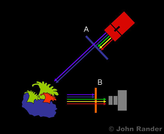
Basically two filters are needed to photograph the fluorescence:
A) a high-pass excitation filter, placed on the flash to excite the fluorescence (removing wavelengths longer than blue), or alternatively you can use UV or deep blue LEDs,
B) a low-pass barrier filter (typically deep yellow) placed on the camera to block the excitation light, while allowing the camera to record the longer wavelength fluorescence.
Choosing the correct barrier filter:
In principle the shorter wavelength cut-off of the barrier filter should match the longer wavelength drop-off of the excitation light. But creative UW lighting objectives may influence your choice of this matching. By allowing a small fraction of the excitation light to pass you can brighten the scene and give a “perspective” which will allow the viewer to better understand the image. However, overdoing this can create some confusion between reflected excitation light and bluish fluorescence, and may even hide weaker fluorescence.A word about aiming lights:
If your filtered flash has a built-in aiming light (located behind the filter), keep in mind that you will not be able to use it to focus a camera equipped with a barrier filter. Alternatively, an external white aiming light should be much weaker than the fluorescence you are photographing, or it should be triggered (with a photodiode) to shut-off when the shutter opens.Fluorescence is common in biology
Chlorophyll fluorescence in plants is well known today; its first observation by H. Kautsky and A. Hirsch dates from 1931. Excited by blue light, plant fluorescence shows peaks in the red near 680 and 700 nm. Measurements of this effect and its changes with time give information on the performance of the plant’s photosynthesis process. The photo (move the mouse over it) illustrates the importance of the low-pass barrier filter when imaging the relatively weak fluorescence (1 to 2% of the absorbed light), excited here with blue LEDs at 470 nm.
Today it is known that some of the fluorescent pigments in corals are forms of the green fluorescent protein (GFP) found in certain medusa. Research in this field and in related applications is in rapid expansion. The 2008 Nobel Prize in chemistry was awarded to Roger Tsien, Martin Chalfie, and Osamu Shimomura for the discovery of GFP and the development of its use as a gene marker.
What is the role of coral fluorescence?
The fluorescent colors might seem fantastic to us, but what use are they for corals? A number of possible roles have been proposed and are actively under study. None have unanimous agreement, often due to conflicting evidence; it’s a complex problem. Most propositions have focused on the possible interplay which might exist between the fluorescent pigments in the coral’s animal tissue and the single-celled algae (called Zooxanthellae colored golden brown, and typically 10µm in diameter) living in a mutually beneficial symbiotic relationship within this tissue. A suggestive piece of evidence along this line is the correlation observed between the amount of green fluorescent protein pigments within the tissue and the amount of blue light naturally reaching the coral in its environment. It has been suggested that the fluorescent pigments might act as a sun-filter to protect the algae, or that the fluorescence might aid photosynthesis in the poorly illuminated depths, or perhaps the fluorescent pigments might play some role in the biochemistry of coral growth. Part of the complexity in finding a clear answer probably comes from the varied localizations of Zooxanthellae in coral tissue depending on different factors and the fact that there are many varieties of these algae. An altogether different approach has suggested that the fluorescence might be a coral defense strategy to change its visual aspect to trick potential predators such as butterfly fish.
Interested readers can explore the publication list on the NightSea website.
Can I see fluorescence while diving underwater?
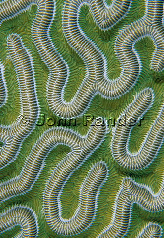
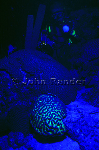
Yes, coral fluorescence can be detected as a curious greenish, orange or even reddish glow or coloration under certain conditions such as under overhangs, or in the late afternoon, just before sunset. It helps to start with subjects which are easy to find. Green fluorescence is generally fairly easy to spot, but if you turn on your (white light) diving light, the effect will disappear. A blue light (filter or blue LED) will allow you to find interesting subjects while night diving. Subjects with weak fluorescence will require adding a special filter to your eyes to block out the blue light, much like police crime investigators looking for evidence.
View my recent images of fluorescence taken in April 2011 on our diving trip to N. Sulawesi in Indonesia: Portfolio Fluorescence
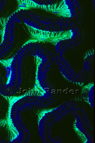
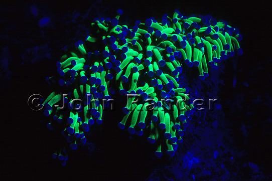
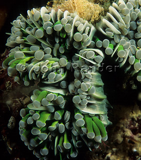
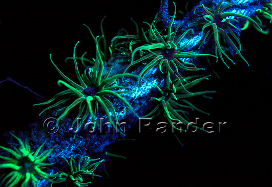
I started using the method on diving trips to Bonaire (March 1998) and Papua New Guinea (May 1998). Since the amount of fluorescence produced is only a fraction of the incident flash, one must generally work in the dark. Night diving with the new light was very exciting, full of unexpected surprises. You will find below some of my images from that period.
Photos: Bonaire (March 1998).
Photos: Kimbe Bay, PNG (May 1998).
Photo: Etang de Maguelone, France (August 1998).
 Top of the page
© John Rander, all rights reserved.